

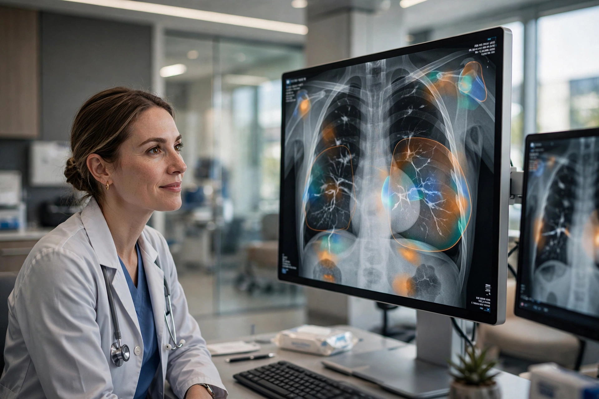
Finding breast cancer early remains one of the most effective ways to improve treatment outcomes, and technology is rapidly changing how that early detection happens. Artificial intelligence is now helping radiologists read mammograms, newer imaging methods are producing clearer pictures of breast tissue, and risk models are identifying women who may benefit from closer monitoring long before any symptom appears.
Reporting by CBS News on new detection technology reflects a broader shift in screening programs: instead of relying on a single image read by a single specialist, clinics are increasingly combining human expertise with software that can flag subtle patterns, prioritize urgent cases and support more personalized screening schedules.
AI as a Second Set of Eyes
AI-assisted mammography is the most closely studied of these advances. Software trained on large collections of earlier scans learns to recognize patterns associated with cancer, including small or developing abnormalities that can be difficult to spot in dense tissue. In practice, the system does not replace the radiologist. It highlights areas worth a closer look, scores cases by risk and helps decide which examinations need the most urgent expert attention.
The strongest evidence so far comes from Sweden’s MASAI trial, a large randomized study involving more than 105,000 women. As summarized in coverage of the trial results, AI-supported screening increased cancer detection by 29 percent without increasing false positives, reduced radiologists’ screen-reading workload by 44 percent, and was associated with 12 percent fewer interval cancers — cancers diagnosed between regular screening rounds. The cancers found with AI support also tended to be detected while they were still relatively small, with fewer invasive and aggressive cases emerging later.
Those interval cancer figures matter because cancers that surface between screenings are often faster-growing and harder to treat. Catching more of them at the screening stage can mean earlier treatment and, in some cases, less invasive procedures. Researchers involved in the trial have stressed, however, that AI tools should be introduced carefully, using tested systems and continuous monitoring of results across different populations.
Better Images, Clearer Answers
Imaging itself is also improving. Digital breast tomosynthesis, commonly called 3D mammography, takes images of the breast from multiple angles and reconstructs them into thin layers. That layered view helps radiologists see through overlapping tissue, a common problem in standard two-dimensional mammograms, and can reduce both missed cancers and unnecessary recalls for additional testing.
For women at higher risk, or whose mammograms remain inconclusive, doctors may also turn to breast MRI and ultrasound as supplementary tools. The trend across all of these methods is the same: gather more information from each examination, and use software to help specialists interpret it consistently. Better imaging also supports the kind of careful, long-term follow-up discussed in our article on maintaining a smooth long-term experience with ongoing care.
Risk Models Are Becoming Personal
Traditional risk assessment relied heavily on age, family history and a small set of known factors. Newer models, including AI-based approaches that analyze the mammogram image itself, can estimate a woman’s risk over the coming years with far more individual detail. A high-risk result does not mean cancer is present. It can instead prompt earlier screening, more frequent examinations or additional imaging, while lower-risk women may safely follow standard schedules.
This personalization is especially relevant for women with dense breasts, where standard mammography performs less well and where risk can also be higher. In the MASAI research, AI support improved sensitivity across age and breast-density groups, including among women with extremely dense tissue, suggesting the tools may help most where conventional screening has traditionally struggled.
What Technology Cannot Do Alone
Specialists caution that detection technology is only one part of the pathway. A suspicious result still requires diagnostic imaging, biopsy and expert review, and access to advanced screening remains uneven between well-funded urban hospitals and smaller clinics. False positives, although not increased in the Swedish trial, can still cause anxiety and extra appointments, which is why accuracy measures such as specificity are watched as closely as detection rates.
Further reporting on the MASAI findings is available from Medical News Today, which details the trial’s results on detection, workload and interval cancers. Ongoing studies of later screening rounds should show whether the early gains translate into long-term improvements in survival.
For now, the direction is clear. Screening is moving from a uniform, one-size-fits-all process toward one guided by sharper images, intelligent software and individual risk. The goal is not simply to find more cancers, but to find the important ones earlier, when treatment has the best chance of success — and technology, used carefully alongside experienced clinicians, is steadily making that possible.
